V Fib Ekg Strip

Ventricular tachycardia a sequence of three pvcs in a row is ventricular tachycardia.
V fib ekg strip. The rate will be 120 200 bpm. Flutter waves are not like well developed p waves with equal pr intervals uniform and consistent see below. The fibrillation is maintained by re entry circuits formed by some of the wavelets. As a result the heart cannot pump blood.
A doctor can identify some types of atrial fibrillation by looking at an electrocardiogram. Symptoms of both afib and vfib are shortness of breath dizziness nausea and chest pain. Ventricular arrhythmias are abnormal heart rhythms originating in the ventricles that are the leading cause of sudden cardiac death. It measures the length of time it takes for the initial impulse to fire at the sinus node and then ends in the contracting of the ventricles.
Which can sort of resemble p waves. The ecg signal strip is a graphic tracing of the electrical activity of the heart. When interpreting a fib on an ekg strip the rhythm will be irregular and not have with sinus rhythm the p waves will be in place with equal pr intervals. The mother rotor then gives rise to propagating unstable daughter wavefronts which results in the chaotic electrical activity seen on the ecg.
Ventricular fibrillation is an emergency condition requiring immediate action. Atrial fibrillation or a fib can lead to fatal heart complications if it reaches a severe enough stage. V fib ventricular fibrillation is an emergency that requires immediate medical attention. Ventricular fibrillation is treated using the left branch of the cardiac arrest algorithm.
It is a frequent cause of sudden cardiac death. Vf can rapidly lead to heart muscle ischemia and there is a high likelihood that it will deteriorate into asystole. When the ventricles handle the pacemaking role they can be observed on ekg tracings. Atrial fibrillation afib and ventricular fibrillation vfib are both a type of abnormal heart rhythm arrhythmia.
Schematic diagram of normal sinus rhythm for a human heart as seen on ecg. Mother rotor mechanism in which a stable re entry circuit is formed the mother rotor. Ventricular tachycardia has two variations monomorphic and. When it occurs the coordinated contraction of the ventricles is replaced rapid chaotic electrical signals.
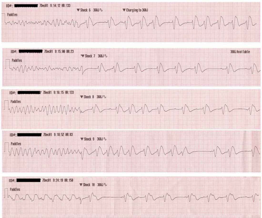
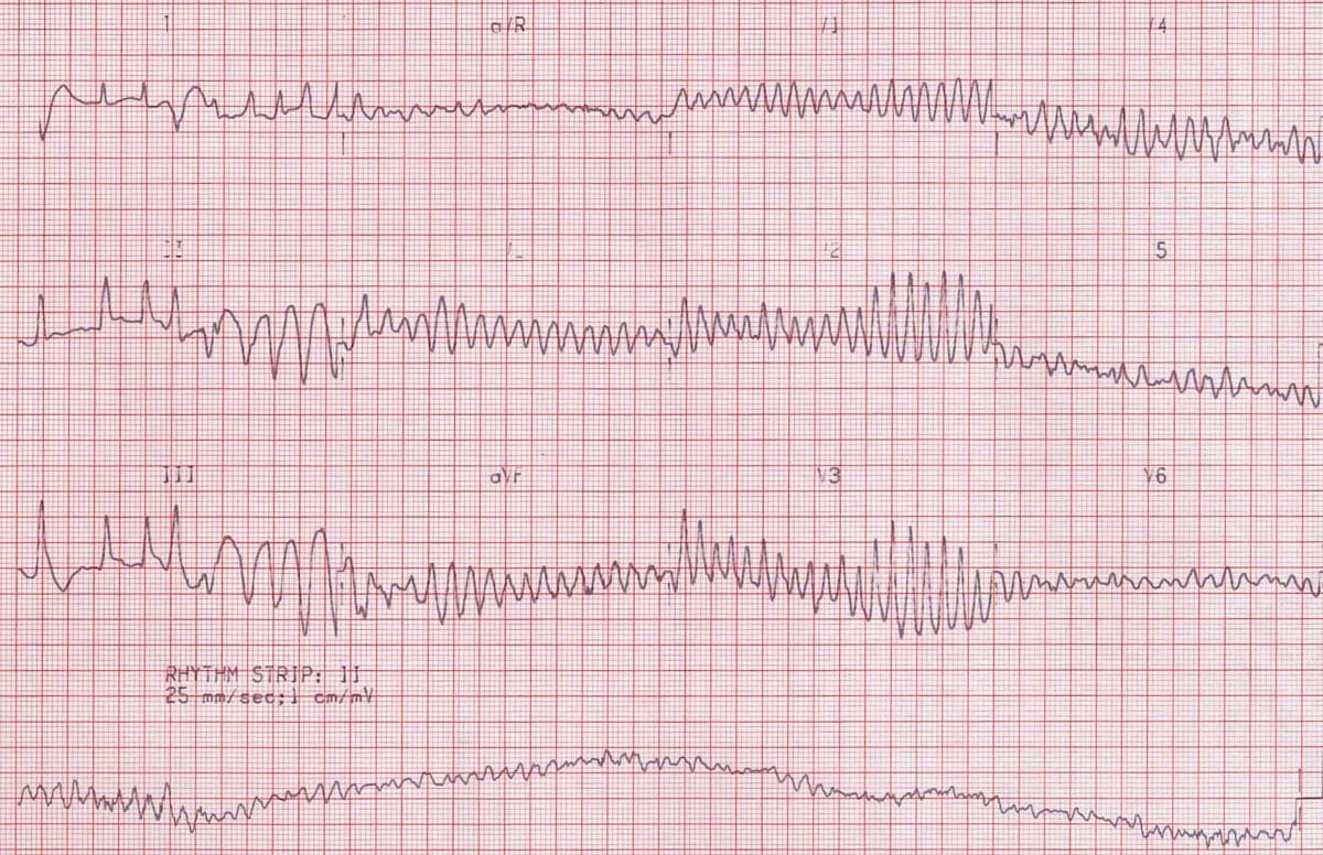
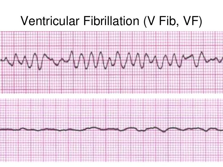
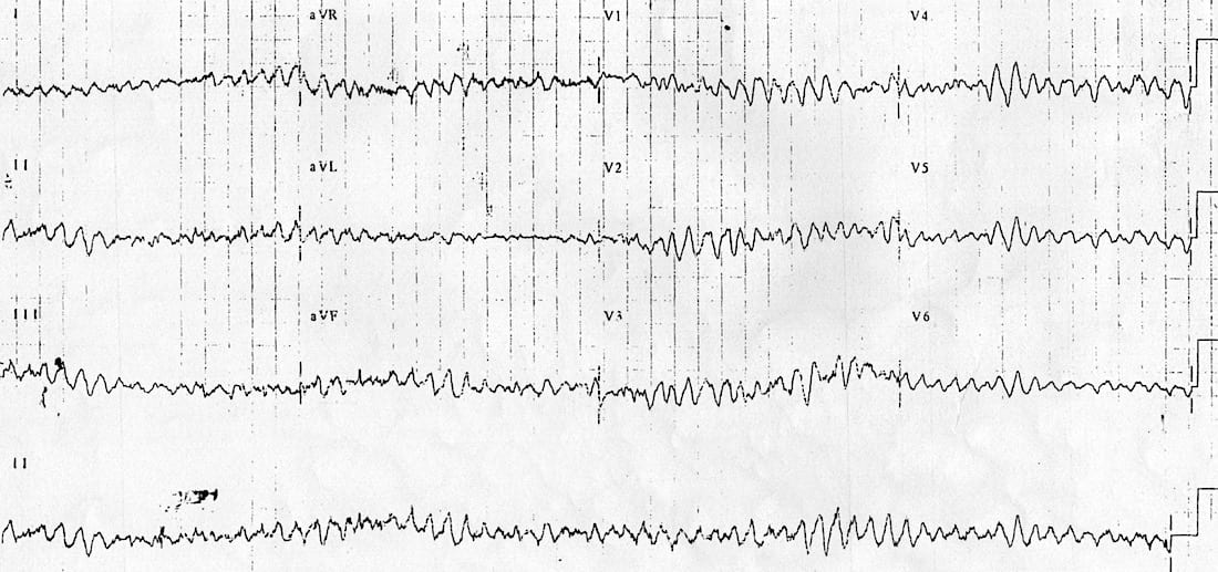
The two images show what ventricular fibrillation will look like on an ekg rhythm strip.


